
At Newcastle Specialist Prosthodontics, we use a range of modern diagnostic tools to support accurate assessment, precise planning and high-quality restorative care.
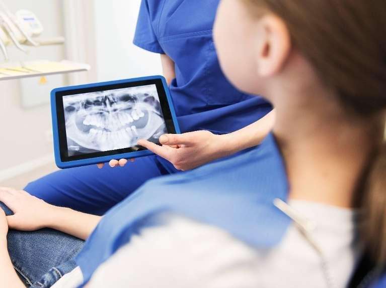
Digital X-Rays
Our practice uses digital intraoral radiography, which provides high-resolution images with significantly lower radiation exposure compared to traditional film.
Types of Radiographs We Take Onsite
Bitewing X-rays:
Taken from inside the mouth, these show the upper and lower back teeth and are ideal for detecting decay between teeth and monitoring existing restorations.
Periapical X-rays:
These capture the entire length of a tooth or dental implant—from crown to root tip—and are used to assess root health, bone levels, infection or structural issues.
When We Refer for Advanced Imaging
Some diagnostic needs require radiographs that cannot be taken in our practice. We work closely with trusted imaging centres for:
OPG (Orthopantomogram):
A full panoramic image showing all teeth, jaws and surrounding structures. This is commonly used for implant planning, assessing bone levels, viewing unerupted or impacted teeth, and assessing the anatomy of the temporomandibular joint (TMJ).
CBCT (Cone Beam CT):
A 3-D scan that provides detailed views of bone, nerves and anatomical landmarks. This advanced imaging is essential for complex implant planning and surgical assessment.
If these images are required, we will organise a referral and review the results with you at your next appointment.
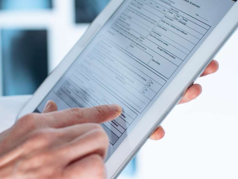
Digital Records
Our practice uses a combination of digital and written records to document your care accurately and securely. Clinical notes and imaging are maintained digitally, allowing us to access information efficiently and monitor changes over time, while some consultation notes and forms are completed on paper where this best supports careful assessment and treatment planning. This blended approach helps ensure continuity of care while allowing flexibility as our systems continue to evolve.
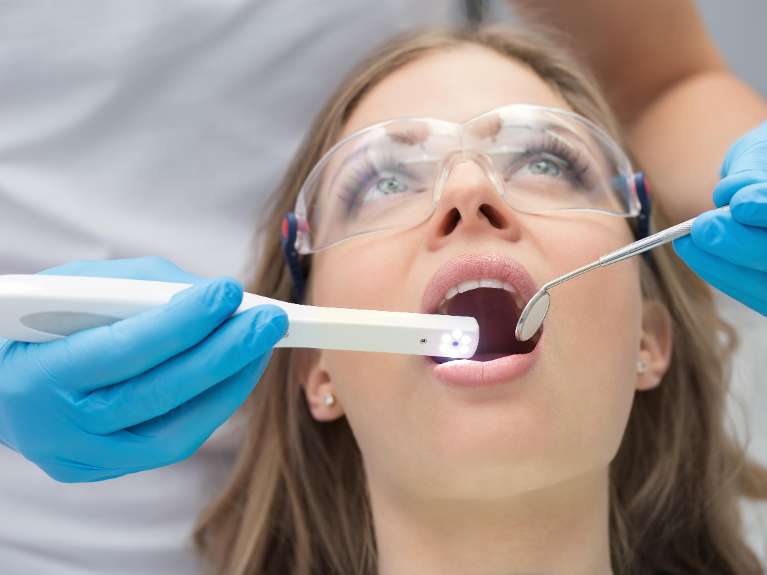
Intraoral Camera
Our intraoral camera allows us to take clear, magnified photographs from inside the mouth. These images are used for:
- Documenting progress and treatment outcomes.
- Monitoring changes over time.
- Explaining conditions or concerns visually.
- Supporting personalised oral hygiene education.
Many patients find it easier to understand their oral health when they can see what we see, making discussions clearer and more collaborative.
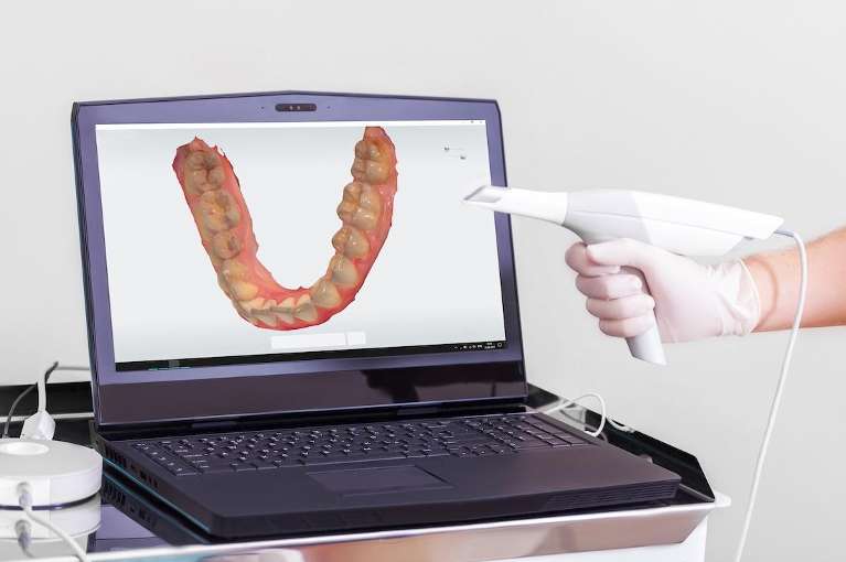
Digital Impressions
We use a digital scanner to capture precise 3-D images of your teeth and bite, replacing the need for conventional impression materials in many situations. Digital impressions offer:
- Enhanced comfort—no impression trays or setting materials.
- Improved accuracy for crowns, bridges, implant restorations and splints.
- Faster communication with dental laboratories.
- Better visualisation during treatment planning.
When We Use Traditional Impressions
Although digital scanning is suitable for most restorative procedures, some diagnostic records still require traditional impressions. These are used to create study models, which we pour in our on-site laboratory. Physical models allow our prosthodontist to examine your bite, function and tooth structure from all angles when planning complex treatment or full-mouth rehabilitation.
Traditional impressions are typically reserved for situations where they provide important diagnostic information—ensuring your treatment is planned with the highest level of accuracy and care.